CASE REPORT
LDT-PPH (PTERODON PUBESCENS) FORMULATION IN THE TOPICAL TREATMENT OF PRESSURE INJURY
FORMULAÇÃO LDT-PPH (PTERODON PUBESCENS) NO TRATAMENTO TÓPICO DE LESÃO POR PRESSÃO
FORMULACIÓN DE LDT-PPH (PTERODON PUBESCENS) EN EL TRATAMIENTO TÓPICO DE LAS LESIONES POR PRESIÓN
https://doi.org/10.31011/reaid-2026-v.100-n.3-art.2747
1Fernanda de Souza Silva
2Luiz Antonio Soares Romeiro
3Priscilla Roberta Silva Rocha
1Enfermeira, mestranda, Faculdade de Ciências da Saúde, Departamento de Enfermagem, Universidade de Brasília, Brasília-DF, Brazil. ORCID: https://orcid.org/0000-0001-5202-0984
2Químico, Doutor, Professor Associado do Departamento de Farmácia, Faculdade de Ciências da Saúde, Universidade de Brasília, Brasília-DF, Brazil. ORCID: https://orcid.org/0000-0001-5679-0820
3Enfermeira, Doutora, Professora Adjunta da Faculdade de Ciências e Tecnologia da Saúde, Universidade de Brasília, Brasília-DF, Brazil. ORCID: https://orcid.org/0000-0002-2058-8548
Corresponding Author
Fernanda de Souza Silva
Instituto Central de Ciências Norte, Campus Universitário Darcy Ribeiro, Asa Norte, Brasília - DF, Brazil. 70910-900 fone: +55 (61) 3107-1711; E-mail: fernanda.de.ssouza@gmail.com
Submission: 08-03-2026
Approval: 12-07-2026
ABSTRACT
Pressure injuries are reportable adverse events and important indicators of healthcare quality. Their management represents a major clinical and organizational challenge due to the complexity of treatment and the associated demands on healthcare resources. This study describes the healing process of a stage IV sacral pressure injury treated with a 1% solution of the hexane extract of Pterodon pubescens fixed oil (LDT-PPH) diluted in the commercial product Dersani®, composed of essential fatty acids, medium-chain triglycerides, and vitamins A and E. An 81-year-old male patient with systemic arterial hypertension, diabetes mellitus, and hypercholesterolemia, and a previous history of acute myocardial infarction, was admitted to a tertiary hospital following an acute ischemic stroke and subsequently developed a sacral pressure injury. Topical treatment with the LDT-PPH solution promoted favorable wound healing, characterized by increased vascularization, stimulation of angiogenesis, and reduction of local inflammation. The wound bed evolved to healthy granulation tissue, followed by satisfactory epithelialization and progressive contraction of the wound edges. These findings suggest that LDT-PPH may represent a promising therapeutic strategy for the management of complex skin lesions, particularly during the inflammatory and proliferative phases of wound healing, due to its potential anti-inflammatory and angiogenic effects.
Keywords: Pressure Ulcer; Wound Healing; Pterodon Pubescens; Phytotherapeutic Drugs.
RESUMO
As lesões por pressão são eventos adversos de notificação obrigatória e constituem importantes indicadores da qualidade da assistência em saúde. Seu manejo representa um desafio clínico e organizacional significativo devido à complexidade do tratamento e às demandas associadas sobre os recursos do sistema de saúde. Este estudo descreve o processo de cicatrização de uma lesão por pressão sacral em estágio IV tratada com uma solução a 1% do extrato hexânico do óleo fixo de Pterodon pubescens (LDT-PPH), diluída no produto comercial Dersani®, composto por ácidos graxos essenciais, triglicerídeos de cadeia média e vitaminas A e E. Trata-se de um paciente do sexo masculino, de 81 anos, com comorbidades como hipertensão arterial sistêmica, diabetes mellitus e hipercolesterolemia, além de histórico prévio de infarto agudo do miocárdio. O paciente foi admitido em um hospital terciário após um acidente vascular cerebral isquêmico agudo e, posteriormente, desenvolveu uma lesão por pressão sacral. O tratamento tópico com a solução LDT-PPH promoveu evolução favorável do processo de cicatrização, caracterizada por aumento da vascularização, estímulo à angiogênese e redução da inflamação local. O leito da ferida evoluiu para a formação de tecido de granulação saudável, seguido de epitelização satisfatória e contração progressiva das bordas da ferida. Esses achados sugerem que o LDT-PPH pode representar uma estratégia terapêutica promissora para o manejo de lesões cutâneas complexas, especialmente durante as fases inflamatória e proliferativa da cicatrização, devido aos seus potenciais efeitos anti-inflamatórios e angiogênicos.
Palavras chaves: Lesão por Pressão; Cicatrização de Feridas; Pterodon Pubescens; Medicamento Fitoterápico.
RESUMEN
Las lesiones por presión son eventos adversos de notificación obligatoria y constituyen importantes indicadores de la calidad de la atención sanitaria. Su manejo representa un desafío clínico y organizacional significativo debido a la complejidad del tratamiento y a las demandas asociadas sobre los recursos del sistema de salud. Este estudio describe el proceso de cicatrización de una lesión por presión sacra en estadio IV tratada con una solución al 1% del extracto hexánico del aceite fijo de Pterodon pubescens (LDT-PPH), diluida en el producto comercial Dersani®, compuesto por ácidos grasos esenciales, triglicéridos de cadena media y vitaminas A y E. Se trata de un paciente masculino de 81 años con comorbilidades que incluían hipertensión arterial sistémica, diabetes mellitus e hipercolesterolemia, además de antecedentes de infarto agudo de miocardio. Fue admitido en un hospital de tercer nivel tras presentar un accidente cerebrovascular isquémico agudo y posteriormente desarrolló una lesión por presión sacra. El tratamiento tópico con la solución LDT-PPH promovió una evolución favorable del proceso de cicatrización, caracterizada por un aumento de la vascularización, estimulación de la angiogénesis y reducción de la inflamación local. El lecho de la herida evolucionó hacia la formación de tejido de granulación sano, seguido de una epitelización satisfactoria y una contracción progresiva de los bordes de la herida. Estos hallazgos sugieren que LDT-PPH puede representar una estrategia terapéutica prometedora para el manejo de lesiones cutáneas complejas, especialmente durante las fases inflamatoria y proliferativa de la cicatrización, debido a sus potenciales efectos antiinflamatorios y angiogénicos.
Palabras clave: Úlcera por Presión; Cicatrización de Heridas; Pterodon Pubescens; Medicamento Fitoterápico.
INTRODUCTION
Pressure injuries (PIs) are adverse events subject to mandatory reporting and are common among critically ill patients and those requiring prolonged hospitalization.(¹˒²) The National Pressure Ulcer Advisory Panel (NPUAP) and the European Pressure Ulcer Advisory Panel (EPUAP) define a PI as localized damage to the skin and/or underlying soft tissue, usually occurring over a bony prominence or associated with the use of a medical device or other artifact. These injuries result from sustained pressure or a combination of pressure and friction and/or shear forces. PIs are classified according to the depth and extent of tissue involvement and may also be classified based on their association with medical devices.(³)
Several intrinsic and extrinsic factors contribute to the development and progression of PIs. Extrinsic factors are related to the patient's environment and include mechanical pressure over bony prominences, with or without shear, as well as moisture, immobility, and microclimate. Intrinsic factors are directly related to the patient and include comorbidities, advanced age, altered level of consciousness, tissue perfusion, nutritional status, and the use of vasoactive drugs.(⁴˒⁵)
PIs represent a global healthcare concern across different levels of care and affect patients of various age groups. Their occurrence is associated with a substantial economic burden on healthcare systems due to increased use of wound care supplies and technologies, medications, surgical interventions, and prolonged hospitalization.(⁶) The prevention and management of PIs are important indicators of healthcare quality and require multidisciplinary strategies targeting modifiable risk factors and appropriate wound management.(⁷) Healthcare-associated PIs may further prolong hospitalization, increase treatment costs, and negatively affect patients' quality of life.(⁶˒⁷)
In recent decades, increasing attention has been given to the use of herbal medicines as potential therapeutic strategies for promoting cutaneous wound healing.(⁸˒⁹) These products have gained relevance in tissue repair research, supporting continued investigation into their therapeutic potential.(⁸⁻¹⁰) Among these medicinal plants, the fixed oil extracted from the seeds of Pterodon pubescens, commonly known as sucupira, sucupira-branca, faveiro, fava-de-sucupira, fava-de-santo-inácio, or sucupira-lisa, has been investigated for its potential effects on wound healing.
In vitro studies and animal models suggest that this oil may promote wound repair through anti-inflammatory, antiedematous, angiogenic, and fibroplasia-related activities. From an economic perspective, the use of herbal medicines derived from abundant native Brazilian plant species may represent a sustainable and potentially cost-effective alternative to conventional approaches, including high-voltage electrical stimulation, laser therapy, negative-pressure wound therapy, and advanced wound dressings such as hydrocolloids, polyurethane foam, and non-adherent dressings.(¹⁰⁻¹⁵)
The fixed oil of Pterodon pubescens seeds contains compounds belonging to the terpene class in its hexane fraction, including vouacapan-derived compounds, geranylgeraniol, and α-humulene. These compounds have been associated with biological activities that may contribute to the wound healing process, with potentially greater therapeutic relevance during the inflammatory and proliferative phases of tissue repair.(¹¹⁻¹⁷)
Pterodon pubescens is a native species of the Brazilian Cerrado and occurs in states including Minas Gerais, São Paulo, Goiás, and Mato Grosso do Sul. Its oil has traditionally been used for treating inflammatory and rheumatic conditions, sore throat, and respiratory disorders.(¹⁸) Given its bioactive compounds, traditional use, and potentially low production cost, further evaluation of its application in wound healing is warranted. Therefore, this case report aimed to describe the healing progression of a stage IV sacral PI treated for 60 days with a 1% solution of the hexane extract of the fixed oil of Pterodon pubescens (LDT-PPH), diluted in the commercial product Dersani® (LDT-PPH 1% + D).
CASE REPORT
In accordance with Resolution No. 466/2012 of the Brazilian National Health Council (CNS/MS), this study was conducted following approval by the Research Ethics Committee of the Faculty of Health Sciences and Technology at the University of Brasília, under report CAEE 84079724.1.0000.8093.
The solution was provided by the Laboratory for the Development of Therapeutic Innovations (LADETER) of the Tropical Medicine Unit, Faculty of Medicine, University of Brasília, which conducts research in the fields of Pharmaceutical and Medicinal Chemistry and Natural Products.
To obtain the LDT-PPH extract, Pterodon pubescens fruits (fava) were dried at room temperature, cut using pruning shears, and extracted in a 2.5-L percolator with a hexane mixture (4 × 24 h), followed by ethanol extraction (3 × 48 h). The hexane extract was obtained by evaporating the solvents under reduced pressure using a rotary evaporator at 35°C, followed by removal of residual solvents under high vacuum and dose standardization.(¹³) The development of pharmaceutical formulations containing Pterodon pubescens fruit extracts was registered in the Brazilian National System for the Management of Genetic Heritage and Associated Traditional Knowledge (SISGEN) under registration number A93F250.
The patient was an 81-year-old man with a history of systemic arterial hypertension (SAH), diabetes mellitus (DM), hypercholesterolemia, and previous acute myocardial infarction (AMI). He was admitted to a private tertiary-care hospital in a city in the state of Minas Gerais, Brazil, following an acute ischemic stroke, presenting with left-sided hemiplegia, dysarthria, and dysphagia, while remaining conscious and oriented. The total hospitalization period, including admission to the intensive care unit (ICU) and subsequent ward stay, was four months.
At hospital admission, the patient's skin was intact. However, during the third week of hospitalization in the ward, he developed a stage 1 sacral pressure injury (erythema). Subsequently, his clinical condition deteriorated, requiring transfer to the ICU, where he remained for 27 days and received invasive mechanical ventilation, vasoactive drugs, and enteral nutrition. Upon discharge from the ICU, the patient had an unstageable sacral pressure injury due to the presence of devitalized tissue, which prevented assessment of the extent and depth of tissue involvement. He also presented with stage 3 pressure injuries in both trochanteric regions.
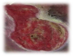
Five months after the ischemic stroke, while receiving home-based care, the patient presented with a stage IV sacral pressure injury consisting predominantly of granulation tissue, with abundant serous exudate (3+/4+), a flat and irregular wound edge, no malodor, and an approximate depth of 3 cm (Figure 1).
Before initiating treatment, photographs of the pressure injury were taken on the first day of the treatment protocol (Day 0; D0), and wound dimensions were measured with a ruler. To systematically assess wound healing progression, the TIMERS framework was used.(¹⁹) The acronym refers to Tissue (management of nonviable tissue), Infection/Inflammation (infection and inflammation control), Moisture (exudate management), Edge (wound-edge management), Regeneration/Repair (tissue regeneration), and Social factors (social factors).(¹⁹) The wound was evaluated according to the TIMERS criteria at each assessment, as presented in the corresponding figures.
Figure 1 - Before treatment with the LDT-PPH 1% + D solution (Day 0; D0). Stage IV sacral pressure injury measuring 12 cm in width (right-to-left), 8 cm in height (sacral-gluteal), and 3 cm in depth. The wound bed consisted predominantly of (T) granulation tissue, with focal areas of liquefactive necrosis in the upper third; (I) no signs of infection; (M) moderate amount of serous exudate (3+/4+); (E) flat, irregular wound edges, with no malodor; and (R) no evidence of wound-edge contraction.
A caregiver was trained by the study nurse (P.R.S.R.) to perform the dressing changes daily using a clean technique. The wound was cleansed daily with approximately 100 mL of sterile 0.9% saline solution by simple jet irrigation. After cleansing, the wound edges were dried, and the LDT-PPH 1% + D solution was applied directly to the entire wound bed. A sterile gauze dressing was used as the secondary dressing. The treatment protocol consisted of applying LDT-PPH 1% + D for 60 days. Dressing changes were performed daily after bathing and, when necessary, for example, in the event of fecal contamination, the dressing was replaced at any time of day using the same technique.
The wound was photographed daily before and after cleaning with 0.9% saline, and the images were sent to the nurse responsible for the study (P.R.S.R.). Subsequent in-person assessments were performed every 15 days under standardized lighting conditions and at the same time of day to minimize potential variations in photographic documentation. Following assessment by the study nurse (P.R.S.R.) and after the patient's family had provided written informed consent, treatment with the 1% LDT-PPH solution in Dersani® (LDT-PPH 1% + D) was initiated.
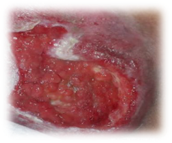
After 13 days of treatment with LDT-PPH 1% + D, a reduction in wound size and depth was observed, accompanied by an increased amount of granulation tissue and progressive wound contraction (Figure 2).
Figure 2 - Day 13 of treatment with LDT-PPH 1% + D solution. Stage IV sacral pressure injury measuring 10 cm in width (right-to-left), 7.5 cm in height (sacral-gluteal), and 2.0 cm in depth. The wound presented (T) predominantly healthy granulation tissue, with progressive filling of the wound bed; (I) no signs of infection; (M) moderate amount of serous exudate (2+/4+); (E) flat, irregular wound edges, with evidence of wound-edge contraction and no malodor; and (R) progressive wound-edge contraction and granulation tissue formation, indicating ongoing epithelialization and tissue regeneration.
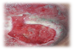
After 28 days of treatment with LDT-PPH 1% + D, a substantial reduction in wound size and depth was observed, accompanied by an increase in granulation tissue and progressive superficialization of the pressure injury (Figure 3). Due to the presence of wound-edge maceration, calcium alginate dressing was added to the treatment protocol to promote exudate absorption and moisture management.
Figure 3 - Day 28 of treatment with LDT-PPH 1% + D solution. Stage IV sacral pressure injury measuring 5 cm in width (right-to-left), 6.5 cm in height (sacral-gluteal), and 0.5 cm in depth. The wound presented (T) healthy granulation tissue; (I) no signs of infection; (M) a small amount of serous exudate (1+/3+), with some maceration of the wound edges; and (E) contracted, flat, regular wound edges, with no malodor.
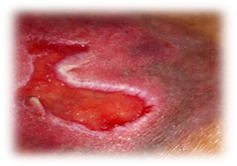
After 60 days of treatment, the pressure injury showed substantial improvement, as illustrated in Figure 4.
Figure 4 - Day 60 of treatment with LDT-PPH 1% + D solution (D60). Sacral pressure injury measuring 3.5 cm in width (right-to-left), 3.45 cm in height (sacral-gluteal), and 0.3 cm in depth. The wound presented (T) healthy granulation tissue; (I) no signs of infection; (M) a small amount of serous exudate (1+/4+); (E) contracted, epithelialized, flat, and regular wound edges, with no malodor; and (R) evidence of epithelialization and tissue regeneration.
During treatment with the LDT-PPH 1% + D solution, progressive healing of the sacral pressure injury was observed based on serial assessments using the TIMERS framework, as illustrated in Figures 2–4. The healing process was characterized by an increase in healthy granulation tissue, progressive superficialization and filling of the wound bed, reduction in wound size, decreased inflammation as evidenced by reduced exudate (1+/4+), improved moisture management with resolution of wound-edge maceration, progressive contraction of the wound edges, and epithelialization of newly formed tissue (Figure 4). Overall, treatment with LDT-PPH 1% + D showed promising results, with the wound demonstrating a continuous and consistent healing trajectory throughout the 60-day follow-up period.
The application of LDT-PPH 1% + D was associated with favorable wound-healing progression according to the parameters assessed. It is noteworthy that the patient presented both intrinsic and extrinsic factors that could delay wound healing and increase the risk of developing new pressure injuries, including diabetes mellitus, glycemic fluctuations despite daily treatment with regular and NPH insulin, impaired mobility, impaired communication, and edema.
The development of pressure injuries is directly associated with increasing patient severity and is influenced by multiple intrinsic and extrinsic factors. Advanced age is associated with reduced skin elasticity and texture, impaired circulation, decreased cellular turnover, and delayed wound healing, as well as diminished peripheral sensitivity, thereby increasing the risk of skin and tissue injury. Conditions such as systemic arterial hypertension, diabetes mellitus, and dyslipidemia may impair the healing process by compromising vascular function. Reduced mobility limits the ability to effectively relieve pressure, while malnutrition may increase the risk of developing pressure injuries by up to fourfold.(⁴˒⁵˒²⁰⁻²⁴)
It is important to highlight the factors associated with the development of pressure injuries (PIs). The use of vasoactive drugs may cause peripheral vasoconstriction, consequently reducing tissue perfusion. Impaired consciousness may compromise the patient's perception of discomfort and, consequently, their ability to respond to prolonged pressure. The use of mechanical ventilation may indicate impaired oxygenation and/or perfusion and, when combined with other risk factors, may further increase the risk of PI development. (⁴˒⁵˒²⁰⁻²⁴)
Moisture is also an important contributing factor, particularly in patients with impaired consciousness who are unable to reposition themselves or communicate their needs to healthcare professionals. Urinary and fecal incontinence further increase skin moisture and exposure to pressure, predisposing the tissue to maceration and breakdown. These conditions may also cause bed linens and clothing to adhere to the skin, thereby intensifying the effects of shear and friction forces, particularly when the head of the bed is elevated above 30° and interface pressure exceeds 32 mmHg. Such forces may result from patient movement or improper positioning. (⁴˒⁵˒²⁰⁻²⁴)
Several of the risk factors described above were present in the clinical condition of the patient reported in this case, including advanced age, chronic diseases, hemiplegia, invasive mechanical ventilation, and the use of vasoactive drugs during ICU admission. Additional risk factors included impaired level of consciousness, reduced mobility, moisture associated with diaper use, shear and friction forces, and ineffective repositioning. The combination of these factors likely increased pressure over bony prominences and contributed to the development and progression of pressure injuries.
Considering the prevention of pressure injuries (PIs), individualized risk stratification should be used to facilitate the early implementation of preventive measures. Among the most used predictive tools is the Braden Scale, which assesses factors associated with the risk of PI development.(⁴˒⁶˒⁷˒²⁰˒²⁴) In the present case, the patient was classified as having a moderate-to-high risk of developing pressure injuries.
Pressure injuries are primarily influenced by two key determinants: the duration and intensity of pressure. In general, stage III and IV pressure injuries are more frequently observed after 30 days of hospitalization and may be associated with increased morbidity and mortality, although earlier-stage injuries may develop within as little as 24 hours.(¹⁰˒²²) Studies have reported that pressure injuries occur most frequently in the sacral, gluteal, and intergluteal regions, as these areas are located over bony prominences and are therefore exposed to greater pressure.(⁴˒⁵˒¹⁰˒²¹˒²²)
Regarding age, studies have reported a higher incidence of pressure injuries among individuals over 60 years of age, with the risk increasing when ICU stays exceed five days. This increased risk is likely related to the cumulative effect of multiple risk factors associated with clinical severity and hemodynamic instability.(⁵˒¹⁰˒²¹⁻²³) These characteristics were present in the patient described in this case, consistent with findings reported in the literature.
Studies have reported that sacral pressure injuries are associated with higher treatment costs and greater therapeutic challenges.(⁶) Therefore, the investigation of accessible and cost-effective alternative approaches that do not compromise treatment effectiveness is relevant for wound care.
According to previous studies, the favorable healing outcomes observed with the hexane extract of Pterodon pubescens may be attributed to its anti-inflammatory, hemostatic, angiogenic, and fibroblast-stimulating (fibroblastic) activities. The extract contains predominantly terpenes, including diterpenes and sesquiterpenes with a vouacapan-type skeleton, such as 6α,7β-dihydroxyvouacapan-17β-oic acid, which are present in the LDT-PPH 1% + D solution.(¹⁰⁻¹⁵)
The anti-inflammatory activity, which has been widely described in the literature, may be associated with the presence of terpenoid derivatives in the hexane fraction, which may contribute to fibroblast stimulation and reduction of edema.(¹²˒¹³) Geranylgeraniol has also been shown to significantly reduce edematous and inflammatory responses.(¹¹˒¹⁵˒²⁶) In addition, α-humulene has been associated with angiogenic activity and, at higher concentrations, may stimulate vascular endothelial growth factor (VEGF), thereby promoting cell migration and proliferation and potentially contributing to wound closure.(¹⁻¹³˒¹⁵˒¹⁶)
The use of LDT-PPH 1% + D solution was associated with favorable wound healing progression and improved vascularization, as evidenced by the development of a bright-red, healthy-appearing wound bed. Subsequently, progressive wound-edge contraction, filling, and superficialization of the lesion were observed, consistent with findings reported in the literature.(²⁷˒²⁸) These findings suggest that topical therapy with LDT-PPH 1% + D may contribute to accelerating the wound-healing process and represents a promising alternative for promoting satisfactory healing in wounds with delayed progression and impaired tissue repair.
Additionally, studies evaluating the safety and metabolic effects of the hexane extract of Pterodon pubescens fixed oil have reported potential beneficial effects on metabolic parameters. In humans, oral administration at a dose of 250 mg/kg was associated with reductions in blood glucose, cholesterol, and triglyceride levels.(²⁹) In animal studies, administration at doses of 100 and 300 mg/kg was associated with reduced body weight, with a hypoglycemic effect observed at the higher dose of 300 mg/kg.(¹⁵) These findings were further supported by subsequent research exploring the use of encapsulated Pterodon pubescens fixed oil as a potential strategy for diabetes management.(³⁰)
Regarding the topical use of LDT-PPH 1% + D in wound care, experimental studies have demonstrated promising results regarding its potential effectiveness. However, clinical evidence in humans remains limited, and further studies are needed to establish its efficacy and safety in the wound-healing process. In this context, controlled clinical trials are warranted to systematically evaluate the therapeutic effects of this formulation across different types of skin wounds. In addition, recent research has explored micro- and nanoencapsulation strategies for Pterodon pubescens oil, which may improve its bioavailability and stability, potentially enhance its therapeutic effects, and contribute to more favorable clinical outcomes in wound management.(²⁶˒³⁰˒³¹)
CONCLUSION
The findings of this case report suggest that the formulation containing Pterodon pubescens may have therapeutic potential in promoting cutaneous wound-healing, possibly through the activity of its terpenoid derivatives, particularly during the inflammatory phase, by modulating excessive inflammation, and the proliferative phase, by promoting angiogenesis. Clinically, favorable wound healing progression was observed, characterized by the development of well-vascularized, bright-red, healthy-appearing granulation tissue, followed by satisfactory epithelialization.
However, important limitations remain regarding the use of Pterodon pubescens as a wound-healing agent. The available scientific evidence is still limited, particularly regarding clinical studies evaluating its topical application in humans. Therefore, further research, preferably well-designed controlled clinical trials, is needed to establish its efficacy and safety in the treatment of cutaneous wounds.
The main limitations of this case report include the limited evidence regarding the topical use of Pterodon pubescens oil in wound healing, both in animal models and in humans. In addition, patient-specific clinical characteristics, such as diabetes mellitus, may have influenced the healing process and limit the generalization of the findings. Further investigations involving patients with different clinical profiles are therefore warranted to better characterize the therapeutic potential of this formulation and strengthen scientific evidence supporting its use in wound care.
1. Pantaleão de Souza TM, Nogueira PC, Conceição de Gouveia Santos VL, Gonçalves Faustino Campanili TC, da Costa Silva Santos RS, da Silva Oliveira EL. Lesão por pressão em pacientes críticos: prevalência e fatores associados. Estima. 2025;22. doi:10.30886/estima.v22.1519_PT
2. Agência Nacional de Vigilância Sanitária (BR). Nota técnica nº 05/2023. Práticas de Segurança do Paciente em Serviços de Saúde: Prevenção de Lesão por Pressão. Brasília: ANVISA; 2023. Disponível em: https://www.gov.br/anvisa/ptbr/centraisdeconteudo/publicacoes/servicosdesaude/notas-tecnicas/notas-tecnicas-vigentes/nota-tecnica-gvims-ggtes-anvisa-no-05-2023-praticas-de-seguranca-do-paciente-em-servicos-de-saude-prevencao-de-lesao-por-pressao
3. National Pressure Injury Advisory Panel; European Pressure Ulcer Advisory Panel; Pan Pacific Pressure Injury Alliance. Pressure ulcers/injuries: definition and etiology. In: Haesler E, editor. Prevention and treatment of pressure ulcers/injuries: clinical practice guideline. 4th ed. 2025. Available from: https://internationalguideline.com
4. Jia YJ, Hu FH, Zhang WQ, Tang W, Ge MW, Shen WQ, et al. Incidence, prevalence and risk factors of device-related pressure injuries in adult intensive care unit: a meta-analysis of 10,084 patients from 11 countries. Wound Repair Regen. 2023;31(5):713-22. doi:10.1111/wrr.13112
5. Teixeira AO, et al. Factors associated with the incidence of pressure wounds in critical patients: a cohort study. Rev Bras Enferm. 2021;75(6):e20210267. doi:10.1590/0034-7167-2021-0267
6. Nascimento FC, et al. Impactos sociais e econômicos das lesões por pressão em unidades de terapia intensiva: uma análise narrativa. OLEL. 2024;22(2):e3375. doi:10.55905/oelv22n2-181
7. Coelho PHS, Oliveira RS, Santos GB. Mapeamento de tecnologias direcionadas à prevenção de lesão por pressão alinhadas às diretrizes internacionais. Cienc Plural. 2023;16(4):1274-91. doi:10.9771/cp.v16i4.50458
8. Carvalho ACB, Perfeito JPS, Britto Vidal Filho W, Silveira D. A decade retrospective and future prospects: how European regulatory alignment shapes Brazil's herbal medicine market. J Ethnopharmacol. 2025;353(Pt B):120404. doi:10.1016/j.jep.2025.120404
9. Cedillo-Cortezano M, Martinez-Cuevas LR, López JAM, Barrera López IL, Escutia-Perez S, Petricevich VL. Use of medicinal plants in the process of wound healing: a literature review. Pharmaceuticals (Basel). 2024;17(3):303. doi:10.3390/ph17030303
10. Bullos BS, et al. Feridas complexas e seus tratamentos alternativos: uma revisão de literatura. Rev Educ Ambient Med. 2022;5:e10010. doi:10.25248/reamed.e10010.2022
11. Silva MC, et al. Acute and topic anti-edematogenic fractions isolated from the seeds of Pterodon pubescens. J Pharm Pharmacol. 2004;56(1):135-41. doi:10.1211/0022357022485
12. Dutra RC, et al. Efeito cicatrizante das sementes de Pterodon emarginatus Vogel em modelos de úlceras dérmicas experimentais em coelhos. Lat Am J Pharm. 2009;28(3):375-82. Disponível em: http://www.latamjpharm.org/trabajos/28/3/LAJOP_28_3_1_8_0O4O1V30HG.pdf
13. Marra AP. Wound healing profile of diabetic rats in the use of LDT-PPH and low level laser therapy [dissertação]. Brasília: Universidade Católica de Brasília; 2010. Disponível em: https://bdtd.ucb.br:8443/jspui/handle/123456789/1124
14. De Araújo LA, et al. Angiogenic activity of sucupira (Pterodon emarginatus) oil. Sci Med. 2015;25(2):ID20351. doi:10.15448/1980-6108.2015.2.20351
15. Basting RT, et al. Pterodon emarginatus: antinociceptive synergistic effect of geranylgeraniol and vouacapans fractions. Rev Bras Farmacogn. 2023;33:856-62. doi:10.1007/s43450-023-00413-7
16. Rocha PRS, Silva FS. Efeitos da fava de sucupira branca (Pterodon pubescens) na cicatrização de feridas: revisão integrativa. Rev Enferm Atual In Derme. 2021;95(36):e21153. doi:10.31011/reaid-2021-v.95-n.36-art.1159
17. Basting RT. Avaliação da associação do extrato bruto diclorometânico de frutos de Pterodon pubescens Benth. e do óleo essencial de Cordia verbenacea DC na atividade antinociceptiva e anti-inflamatória [tese]. Campinas: Universidade Estadual de Campinas; 2018. Disponível em: http://repositório.unicamp.br/jspui/handle/REPOSIP/333660
18. Souza VHDS, et al. Evaluation of non-clinical toxicity of extract and vouacapans from fruits of Pterodon pubescens Benth. Drug Chem Toxicol. 2022;45(3):1325-33. doi:10.1080/01480545.2020.1822858
19. Lumbers M. TIMERS: undertaking wound assessment in the community. Br J Community Nurs. 2019;24(Suppl 12):S22-5. doi:10.12968/bjcn.2019.24.Sup12.S22
20. Itoh S, Yokota S, Yotsu RR, et al. Factors associated with pressure ulcers among middle-old and oldest-old inpatients in an acute care hospital. Sci Rep. 2025;15:23538. doi:10.1038/s41598-025-03937-1
21. Alderden J, Brooks KR, Kennerly SM, Yap TL, Dworak E, Cox J. Risk factors for pressure injuries in critical care patients: an updated systematic review. Int J Nurs Stud. 2025;169:105127. doi:10.1016/j.ijnurstu.2025.105127
22. Chung ML, Widdel M, Kirchhoff J, et al. Risk factors for pressure injuries in adult patients: a narrative synthesis. Int J Environ Res Public Health. 2022;19(2):761. doi:10.3390/ijerph19020761
23. Zhang XH, Fan X, Cai D, Yu JA. Nursing challenges in pressure injury management: global prevalence patterns and projected care demands in aging populations. J Adv Nurs. 2023;ahead of print. doi:10.1111/jan.70121
24. Munoz DCN, et al. Malnutrition and pressure injury risk in vulnerable populations: application of the 2019 International Clinical Practice Guideline. Adv Skin Wound Care. 2022;35(3):156-65. doi:10.1097/01.ASW.0000816332.60024.05
25. Wang I, Walker RM, Gillespie BM, Scott I, Sugathapala RDUP, Chaboyer W. Risk factors predicting hospital-acquired pressure injury in adult patients: an overview of reviews. Int J Nurs Stud. 2024;150:104642. doi:10.1016/j.ijnurstu.2023.104642
26. De Alcantara J, et al. Recent progress in micro and nano-encapsulation of bioactive derivatives of the Brazilian genus Pterodon. Biomed Pharmacother. 2021;143:112137. doi:10.1016/j.biopha.2021.112137
27. Brito AB, Rodrigues V. Análise comparativa de perfil cicatricial utilizando extrato Sucupira Branca (Pterodon pubescens) em paciente com diabetes mellitus tipo II [iniciação científica]. Brasília: Centro Universitário de Brasília; 2022. doi:10.5102/pic.n0.2021.8917
28. Lopes DJ, et al. Investigação do potencial cicatricial da semente de Pterodon emarginatus Vogel. CBE. 2023. Disponível em: https://anais.sobest.com.br/cbe/article/view/854
29. Hoscheid J, et al. Inhibitory effect of the hexane fraction of the ethanolic extract of the fruits of Pterodon pubescens Benth in acute and chronic inflammation. Evid Based Complement Alternat Med. 2013;2013:272795. doi:10.1155/2013/272795
30. Vieira R, et al. Sucupira oil-loaded nanostructured lipid carriers (NLC): lipid screening, factorial design, release profile, and cytotoxicity. Molecules. 2020;25(3):685. doi:10.3390/molecules25030685
31. Giarola LR, et al. Pterodon pubescens Benth (sucupira) microencapsulation influence on formulation stability outcome compared to non-encapsulated extract. J Drug Deliv Sci Technol. 2022;67:102875. doi:10.1016/j.jddst.2021.102875
Funding and Acknowledgments
Acknowledgments to the Therapeutic Innovation Development Laboratory (LADETER) at the Center for Tropical Medicine, Faculty of Medicine, University of Brasília, particularly to the laboratory coordinator, Prof. Dr. Luiz Antônio Soares Romeiro, for his collaboration and for providing supplies for the research.
Data availability statement
No datasets were generated in this study. The information presented is described in the body of the article.
Conflict of interest statement
None to declare.
Authorship criteria
Fernanda de Souza Silva and Priscilla Roberta da Silva Rocha: 1. contributed substantially to the conception and/or planning of the study; 2. to the acquisition, analysis, and/or interpretation of data; and 3. to the drafting and/or critical revision and final approval of the published version.
Luiz Antônio Soares Romeiro contributed by providing the formulation used in the research, as well as to the drafting and/or critical revision and final approval of the published version.
Scientific Editor: Ítalo Arão Pereira Ribeiro. Orcid: https://orcid.org/0000-0003-0778-1447
Rev Enferm Atual In Derme 2026;100(3): e026088